Pathologists have a significant impact on the accuracy of clinical diagnoses. Patient management decisions are dependent upon the accuracy of the reports generated by pathologists, which guide clinicians in ensuring optimal therapeutic plans are formulated.
While pattern recognition by pathologists is subject to intra- and inter-observer variations, this variability can be amplified by the training biases established during residency (2). Between institutions and even among faculty members there may exist inter-observer variability, which can be passed on to students as a result of non-standardized education about diagnostic criteria.
Standardized pathology education is crucial in reducing interobserver variability throughout a pathologist's career, leading to more accurate diagnosis and better patient care. How can faculty and institutions ensure they are providing a standardized educational experience and reducing instances of interobserver variability down the line?
How residents develop diagnostic expertise
To develop diagnostic expertise, pathology residents require long periods of training to expose them to a sufficiently large number of cases, including a wide variety of less common and unusual lesions (5).
In a study of diagnostic accuracy, differences between “novices” (third-year medical students who had recently completed the required second-year course material in pathology), “intermediates” (second- and third-year residents in pathology, who had completed at least one year of surgical pathology), and “experts” (board-certified pathologists, many with special expertise in breast pathology, with an average of 26.5 years of training and practice experience), found that “novices” performed at 2.5% accuracy on average, “intermediates” performed at 40% accuracy, and experts at 78% accuracy (3).
In another study about pathologist interpretation, eye movements, viewer tool behavior (zooming, panning), and interpretation time were tracked using virtual slides of breast lesions. Pathologist experience levels, case consensus diagnosis, case difficulty, eye fixation durations, and the extent to which pathologists' eyes fixated within versus outside of diagnostic regions of interest, all independently or collectively predicted diagnostic accuracy. For example, pathologists with relatively low expertise in interpreting breast pathology were more likely to fixate on and return to diagnostically irrelevant regions of interest relative to experts (6).
Tracking individual residents over time in their learning process shows that with each successive year of experience, the residents' visual search patterns change, including taking significantly less time to view an individual slide and decide where to zoom, significantly fewer fixations are generated overall, and there is less examination of diagnostically irrelevant areas. The more hands-on experience and practice a resident has, the more efficient their search becomes (7).
That is why residency plays a critical role in developing a pathologists understanding and experience. What they learn during this time serves as the foundation of their knowledge, and carries with them throughout their career.
What leads to non-standardized education?
Limited access to rare cases
One factor at play is the way most residency rotations are organized. These tend to be subspecialty based rotations over short periods of time. While such a system will usually expose trainees to commonly occurring lesions, uncommon and rare lesions are often not experienced during such rotations.
Residents can make up for this by reviewing teaching collections of slides. These slides, however, are not annotated, so unless they are reviewed with teaching faculty using double-headed microscopes, may lead to inadequate training due to the defective visual search patterns described above. Such teaching collections are built on the interests of the teaching faculty and are rarely comprehensive.
Faculty workload and time constraints
Another factor is the faculty themselves. Teaching faculty are often overloaded with diagnostic service work. The workload of pathologists is not only influenced by the numbers of cases, specimens, and slides to be interpreted, but also by the degree of effort required to render a diagnosis. A study showed that the evolution of diagnostic procedures over the last decade has resulted in a 60% increase in slide numbers per case, doubling of immunohistochemistry procedures, and more than tripling of molecular analyses but without commensurate adjustment of pathology manpower (1).
However, the nature of pathology education necessitates that senior pathologists teach residents one-to-one and in small groups. As such, there is a burden on faculty to allocate adequate time for teaching residents, participating in mandatory quality assurance activities and professional development programs, all while maintaining their own clinical workflow.
Analog teaching methods
The analog methods used to teach residents restricts their ability for self-study as they need to be in the laboratory to access teaching material (glass slides) and microscopes. Residents are required to share teaching slides which is particularly difficult for small biopsies or rare conditions where sufficient numbers of slides cannot be generated due to tissue depletion. In addition to this, glass slides lack annotation of key features important in pattern recognition which cannot be replaced by descriptive text.
In addition, teaching collections between institutions vary greatly. Some faculty may have access to rare or uncommon slides, while others may not. The learning opportunities are thus greatly reduced for trainees that do not have access to these cases. Non-standardized teaching sets between institutions and faculty creates a knowledge and experience imbalance for future pathologists.
Training future pathologists also requires faculty members to supervise several residents and test their ability to recognize disease markers in tissue using a microscope – a time consuming, laborious and analog process. To achieve this, educators must spend a significant amount of one-on-one time with residents using a double-headed microscope.
Flight simulators and pilot licensing – lessons for maintenance of competence for pathologists
An excerpt from the International Air Transport Association “Guidance Material and Best Practices for Pilot Aptitude Testing”:
Human error consistently accounts for the majority of all aircraft accidents therefore investment in human performance is essential. Investment in the selection process is the foundation of any effort to achieve a quantifiable reduction in accident rates. Human performance is becoming even more relevant because of accelerating innovation and technological change as a result of research and development in the aviation industry. This process of continuous improvement induces changes in the job requirements for pilots and consequently drives the need for continuous adaptation of pilot behavior and airline training methodologies. We cannot rely solely on past experience, because current and future generations of pilot candidates are also evolving. The implementation of a robust and transparent aptitude testing system process will allow an airline to ascertain whether or not a pilot is willing and capable of accepting the inevitable pressures of life-long learning, and training and checking, which are prerequisites for flying technologically advanced aircraft.
The challenges are similar in both aviation and pathology due to acceleration of new knowledge, innovation and technological change influenced by research and development. Pilots and pathologists need to keep on top of such changes in their fields, to ensure passenger and patient safety.
Of the large number of parameters used by the aviation industry to ensure pilots are competent and safe is their performance in full flight simulators. Flight checks in full-flight simulators are commonly used to test flying skills of entry pilots, first officers and captains.
Is there a similar tool for pathology education to standardize education and reduce interobserver variability?
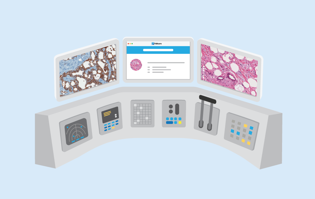
Pathcore Scholar - the diagnostic simulator for anatomic pathology
Pathcore Scholar is designed to simulate a pathologist’s daily workflow including initial consultation. interpretation of whole slide images, ancillary testing, diagnosis, to sign-out. Immediate feedback is provided indicating accuracy of diagnosis, and the appropriateness of choosing one or more biomarker tests.
Within Scholar, instructors can draw from repositories of virtual slides to create precise, standardized case sets. On those virtual slides, they can create detailed, ever-lasting annotations to ensure comprehensive instruction without direct supervision. Trainees can use a WSI viewer - with virtual tests - to inform a virtual diagnosis in a patient-safe environment. Feedback can be immediate and automated, tracking the student’s steps and assessing their journey (including all the tests they ran), not just their diagnosis. Results are logged and tracked, so instructors can refer to accuracy trends overtime. Lastly, each diagnostic simulation and case set can be authored, saved, and shared.
Every step of a pathologist’s daily workflow is accessible, wherever the internet is, on any internet-enabled device. Scholar’s web interface makes collaborating and sharing teaching materials easy, but it also expands learning opportunities far beyond the lab’s doors. Self-study can flourish, with students practicing diagnosis wherever they can find a stable connection. Faculty, meanwhile, can use the extra time self-study affords to refine lab hours or focus on their own clinical caseload.
Collaboration between institutions can further expand the spectrum of disease representation and standardization, ensuring that future pathologists are able to handle any level of complexity no matter where they work.
Learn how Pathcore’s first-of-its-kind diagnostic simulator for pathology education can transform your educational workflows. Schedule a discovery call with our product experts to learn how PathcoreScholar can work for you!
Sources:
1. Warth A, Stenzinger A, Andrulis M, Schlake W, Kempny G, Schirmacher P, et al. Individualized medicine and demographic change as determining workload factors in pathology: quo vadis? Virchows Arch. 2016;468(1):101-8.
2. Raab SS, Grzybicki DM, Janosky JE, Zarbo RJ, Meier FA, Jensen C, et al. Clinical impact and frequency of anatomic pathology errors in cancer diagnoses. Cancer. 2005;104(10):2205-13.
3. Jackson SL, Frederick PD, Pepe MS, Nelson HD, Weaver DL, Allison KH, et al. Diagnostic Reproducibility: What Happens When the Same Pathologist Interprets the Same Breast Biopsy Specimen at Two Points in Time? Ann Surg Oncol. 2017;24(5):1234-41.
4. Zarbo RJ, Meier FA, Raab SS. Error detection in anatomic pathology. Arch Pathol Lab Med. 2005;129(10):1237-45.
5. Crowley RS, Naus GJ, Stewart J, Friedman CP. Development of visual diagnostic expertise in pathology -- an information-processing study. J Am Med Inform Assoc. 2003;10(1):39-51.
6. Brunyé TT, Carney PA, Allison KH, Shapiro LG, Weaver DL, Elmore JG. Eye movements as an index of pathologist visual expertise: a pilot study. PLoS One. 2014;9(8):e103447.
7. Krupinski EA, Graham AR, Weinstein RS. Characterizing the development of visual search expertise in pathology residents viewing whole slide images. Hum Pathol. 2013;44(3):357-64.